What Is Melanoma?
 The most dangerous form of skin cancer, these cancerous growths develop when unrepaired DNA damage to skin cells (most often caused by ultraviolet radiation from sunshine or tanning beds) triggers mutations (genetic defects) that lead the skin cells to multiply rapidly and form malignant tumors. These tumors originate in the pigment-producing melanocytes in the basal layer of the epidermis. Melanomas often resemble moles; some develop from moles. The majority of melanomas are black or brown, but they can also be skin-colored, pink, red, purple, blue or white.
Melanoma is caused mainly by intense, occasional UV exposure (frequently leading to sunburn), especially in those who are genetically predisposed to the disease. Melanoma kills an estimated 8,790 people in the US annually.
If melanoma is recognized and treated early, it is almost always curable, but if it is not, the cancer can advance and spread to other parts of the body, where it becomes hard to treat and can be fatal. While it is not the most common of the skin cancers, it causes the most deaths. The American Cancer Society estimates that at present, about 120,000 new cases of melanoma in the US are diagnosed in a year. In 2010, about 68,130 of these were invasive melanomas, with about 38,870 in males and 29,260 in women.
Warning Signs: The ABCDEs of Melanoma
Moles, brown spots and growths on the skin are usually harmless – but not always. Anyone who has more than 100 moles is at greater risk for melanoma. The first signs can appear in one or more atypical moles. That's why it's so important to get to know your skin very well and to recognize any changes in the moles on your body. Look for the ABCDE signs of melanoma, and if you see one or more, make an appointment with a physician immediately.
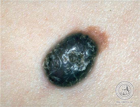
What does a melanoma look like?
All melanomas do not look the same, and there are several different types. The ABCD system (below) tells you some of the things to look out for. A melanoma may show one or more of the following features:
- Asymmetry — the two halves of the area differ in their shape.
- Border — the edges of the area may be irregular or blurred, and sometimes show notches.
- Colour — this may be uneven. Different shades of black, brown and pink may be seen.
- Diameter - most melanomas are at least 6 mm. in diameter.
Melanomas can appear on any part of the skin but they are most common in men on the body, and in women on the legs.
Warning Signs and Images
Even if you have carefully practiced sun safety all summer, it's important to continue being vigilant about your skin in fall, winter, and beyond. Throughout the year, you should examine your skin head to toe once a month, looking for any suspicious lesions. Self-exams can help you identify potential skin cancers early, when they can almost always be completely cured.
First, for a successful self-exam, you obviously need to know what you're looking for. As a general rule, to spot either melanomas or non-melanoma skin cancers (such as basal cell carcinoma and squamous cell carcinoma), take note of any new moles or growths, and any existing growths that begin to grow or change significantly in any other way. Lesions that change, itch, bleed, or don't heal are also alarm signals.
It is so vital to catch melanoma, the deadliest form of skin cancer, early that physicians have developed two specific strategies for early recognition of the disease: the ABCDEs and the Ugly Duckling sign.
Causes and Risk Factors
Am I at Risk?
Everyone is at some risk for melanoma, but increased risk depends on several factors: sun exposure, number of moles on the skin, skin type and family history (genetics).
Sun Exposure
Both UVA and UVB rays are dangerous to the skin, and can induce skin cancer, including melanoma. Blistering sunburns in early childhood increase risk, but cumulative exposure also may be a factor. People who live in locations that have more sunlight – like Florida, Hawaii, and Australia – develop more skin cancers. Avoid using a tanning booth or tanning bed, since it increases your exposure to UV rays, raising your risk of developing melanoma and other skin cancers.
Moles
There are two kinds of moles: normal moles – the small brown blemishes, growths, or "beauty marks" that appear in the first few decades of life in almost everyone – and atypical moles, also known as dysplastic nevi. Atypical moles can be precursors to melanoma, and having them puts you at increased risk of melanoma. But regardless of type, the more moles you have, the greater your risk for melanoma.
Skin Type
- As with all skin cancers, people with fairer skin (who often have lighter hair and eye color as well) are at increased risk.
- Personal History
- Once you have had melanoma, you run an increased chance of recurrence. People who have or had basal cell carcinoma or squamous cell carcinoma are also at increased risk for developing melanoma.
Weakened Immune System
Compromised immune systems as the result of chemotherapy, an organ transplant, excessive sun exposure, and diseases such as HIV/AIDS or lymphoma can increase your risk of melanoma.
Family History
Heredity plays a major role in melanoma. About one in every 10 patients diagnosed with the disease has a family member with a history of melanoma. If your mother, father, siblings or children have had a melanoma, you are in a melanoma-prone family. Each person with a first-degree relative diagnosed with melanoma has a 50 percent greater chance of developing the disease than people who do not have a family history of the disease.
Close Relatives Examined
When this skin cancer is diagnosed, it is standard practice for physicians to recommend that close relatives be examined immediately for melanoma and for the presence of unusual or atypical moles. These moles are also called "dysplastic nevi."
Family Syndrome
When atypical moles are found in an individual belonging to a melanoma family, the condition is known as FAMMM, standing for Familial Atypical Multiple Mole Melanoma Syndrome. People with this syndrome are at the greatest risk of developing melanoma. In contrast, a research study found that those family members who did not have atypical moles were much less likely to develop melanoma.
Genetic Risk Factors
A mutation (alteration) in a recently discovered gene, BRAF, may play a part in causing melanoma. In one study, this mutated gene was found in two-thirds of the melanoma cells analyzed. BRAF is called a "switch" gene, because it turns on to allow the cells to grow and divide. Mutations in this gene can lead to uncontrolled cell growth and cancer. The discovery of BRAF was an exciting research breakthrough, and with the development of the experimental therapy PLX-4032 (a.k.a. Zelboraf) to inhibit BRAF, physicians and patients are just starting to reap some rewards. Ultimately, increasing understanding of the BRAF gene could lead to the development of new diagnostic tools as well as new and improved drug therapies.
The mutations most commonly seen in familial melanoma occur in another gene, p53. When this gene is in its normal state, it functions as a tumor suppressor, giving damaged cells time to repair themselves without progressing to cancer. However, when the gene is altered, it becomes unable to perform this function, and cancer can result.
A number of gene mutations in addition to p53 and BRAF have been associated with familial melanoma, notably the CDKN2A (cyclin-dependent kinase inhibitor 2A) gene. In the future, families might be screened to identify those members who are carrying a defective gene. If, as a result, they become particulary viligant in watching their moles and having regular total-body skin examinations, a melanoma will be detected at its earliest stage, when the chances of a cure are excellent. In fact, testing is now commercially available for the presence or absence of the CDKN2A gene, but the consensus of melanoma experts is that genetic testing is not yet warranted for most people and should be done only in the context of clinical trials.
Moles in an Active Stage
Moles in people belonging to melanoma-prone families are subject to change at certain times of life. They may get larger or show alterations in color or elevation, so for those periods, they are described as being active. While the reasons for these changes are not fully known, there could be a hormonal component: Moles are more active at puberty and during pregnancy. Many – but not all – physicians advise high-risk individuals not to take hormonal medications, such as oral contraceptives or hormone replacement therapy.
Examination Scheduling
Individuals with atypical mole syndrome can improve their chances of early detection by increasing the frequency of skin self-examination and by visiting a physician more often for a full-body skin exam. The clinician may take photographs to document whether there are new moles or changes in older ones.
Children: A Special Case
Children in melanoma-prone families need special care, because familial melanoma is likely to make its appearance early in life. Even though these cancers usually do not appear until after adolescence, they may arise in much younger children who have a family history of melanoma. Most physicians, therefore, advise parents to make a point of studying a child's skin frequently from infancy on.
Physician examination in these families should start at the age of 10 and continue on a twice-a-year basis thereafter. Particular care should be taken at puberty and during adolescence when hormonal changes activate the moles. Here is some encouraging news: Because melanoma families are on the lookout for the disease and seek professional consultation early, the survival rate for familial melanoma is even higher than that for non-familial melanomas.
Prevention Guidelines
Since its inception in 1979, The Skin Cancer Foundation has always recommended using a sunscreen with an SPF 15 or higher as one important part of a complete sun protection regimen. Sunscreen alone is not enough, however. Read our full list of skin cancer prevention tips.
- Seek the shade, especially between 10 AM and 4 PM.
- Do not burn.
- Avoid tanning and UV tanning booths.
- Cover up with clothing, including a broad-brimmed hat and UV-blocking sunglasses.
- Use a broad spectrum (UVA/UVB) sunscreen with an SPF of 15 or higher every day. For extended outdoor activity, use a water-resistant, broad spectrum (UVA/UVB)
sunscreen with an SPF of 30 or higher. - Apply 1 ounce (2 tablespoons) of sunscreen to your entire body 30 minutes before going outside. Reapply every two hours or immediately after swimming or excessive sweating.
- Keep newborns out of the sun. Sunscreens should be used on babies over the age of six months.
- Examine your skin head-to-toe every month.
- See your physician every year for a professional skin exam.
Types of Melanoma
The Four Basic Types Melanomas fall into four basic categories. Three of them begin in situ – meaning they occupy only the top layers of the skin – and sometimes become invasive; the fourth is invasive from the start. Invasive melanomas are more serious, as they have penetrated deeper into the skin and may have spread to other areas of the body.
Superficial spreading melanoma is by far the most common type, accounting for about 70 percent of all cases. This is the one most often seen in young people. As the name suggests, this melanoma grows along the top layer of the skin for a fairly long time before penetrating more deeply.
The first sign is the appearance of a flat or slightly raised discolored patch that has irregular borders and is somewhat asymmetrical in form. The color varies, and you may see areas of tan, brown, black, red, blue or white. This type of melanoma can occur in a previously benign mole. The melanoma can be found almost anywhere on the body, but is most likely to occur on the trunk in men, the legs in women, and the upper back in both.
Lentigo maligna is similar to the superficial spreading type, as it also remains close to the skin surface for quite a while, and usually appears as a flat or mildly elevated mottled tan, brown or dark brown discoloration. This type of in situ melanoma is found most often in the elderly, arising on chronically sun-exposed, damaged skin on the face, ears, arms and upper trunk. Lentigo maligna is the most common form of melanoma in Hawaii. When this cancer becomes invasive, it is referred to as lentigo maligna melanoma.
Acral lentiginous melanoma also spreads superficially before penetrating more deeply. It is quite different from the others, though, as it usually appears as a black or brown discoloration under the nails or on the soles of the feet or palms of the hands. This type of melanoma is sometimes found on dark-skinned people, and can often advance more quickly than superficial spreading melanoma and lentigo maligna. It is the most common melanoma in African-Americans and Asians, and the least common among Caucasians.
Nodular melanoma is usually invasive at the time it is first diagnosed. The malignancy is recognized when it becomes a bump. It is usually black, but occasionally is blue, gray, white, brown, tan, red or skin tone.
The most frequent locations are the trunk, legs, and arms, mainly of elderly people, as well as the scalp in men. This is the most aggressive of the melanomas, and is found in 10 to 15 percent of cases.
The Stages of Melanoma
Once the type of melanoma has been established, the next step is to classify the disease as to its degree of severity.
Classifications for melanomas are called stages. The stage refers to the thickness, depth of penetration, and the degree to which the melanoma has spread. The staging is used to determine treatment.
Early melanomas (Stages I and II) are localized, and more advanced melanomas (Stages III and IV) have spread (metastasized) to other parts of the body. There are also subdivisions within stages.
Treatments
Surgical Techniques Improve
The first step in treatment is the removal of the melanoma, and the standard method of doing this is by surgical excision (cutting it out). Surgery has made great advances in the past decade, and much less tissue is removed than was customary in the past. Patients do just as well after the lesser surgery, which is easier to tolerate and produces a smaller scar. Surgical excision is also called resection, and the borders of the entire area excised are known as the margins.
Outpatient/Office Surgery
In most cases, the surgery for thin melanomas can be done in the doctor’s office or as an outpatient procedure under local anesthesia. Stitches (sutures) remain in place for one to two weeks, and most patients are advised to avoid heavy exercise during this time. Scars are usually small and improve over time.
Discolorations and areas that are depressed or raised following the surgery can be concealed with cosmetics specially formulated to provide camouflage. If the melanoma is larger and requires more extensive surgery, a better cosmetic appearance can be obtained with flaps made from skin near the tumor, or with grafts of skin taken from another part of the body. For grafting, the skin is removed from areas that are normally or easily covered with clothing.
There is now a trend towards performing sentinel node biopsy and tumor removal surgery at the same time, provided the tumor is 1 mm or more thick. When the procedures are combined in this way, the patient is spared an extra visit.
Setting The Margins
In the new approach to surgery, much less of the normal skin around the tumor is removed and the margins, therefore, are much narrower than they ever were before. This spares significant amounts of tissue and reduces the need for postoperative cosmetic reconstructive surgery.
Most US surgeons today follow the guidelines recommended by the National Institutes of Health and the American Academy of Dermatology Task Force on Cutaneous Melanoma.
When there is an in situ melanoma, the surgeon excises 0.5-1 centimeter of the normal skin surrounding the tumor and takes off the skin layers
down to the fat.
- In removing an invasive melanoma that is 1 mm or less in Breslow’s thickness, the margins of surrounding skin are extended to 1 cm and the excision goes through all skin layers and down to the fascia (the layer of tissue covering the muscles).
- If the melanoma is 1.01 to 2 mm thick, a margin of 1-2 cm is taken.
- If the melanoma is 2.01 mm thick or greater, a margin of 2 cm is taken.
These margins all fall within the range of what is called “narrow” excision. When you consider that until recently, margins of 3 to 5 cm (wide excision) were standard, even for comparatively thin tumors, you can see how dramatically surgery has changed for the better. Physicians now know that even when melanomas have reached a thickness of 4 mm or more, increasing the margins beyond 2 cm does not increase survival.
Mohs Micrographic Surgery
In recent years, Mohs Micrographic Surgery, which many physicians consider the most effective technique for removing basal cell and squamous cell carcinomas (the two most common skin cancers), is being increasingly used as an alternative to standard excision for certain melanomas. In this technique, one thin layer of tissue is removed at a time, and as each layer is removed, its margins are studied under the microscope for the presence of cancer cells. If the margins are cancer-free, the surgery is ended.
If not, more tissue is removed, and this procedure is repeated until the margins of the final tissue examined are clear of cancer. Mohs surgery thus can eliminate the guesswork in the removal of skin cancers and pinpoint the cancer’s location when it is invisible to the naked eye.
Mohs surgery differs from other techniques since the microscopic examination of all excised tissues during the surgery eliminates the need to “estimate” how far out or deep the roots of the skin cancer go. This allows the Mohs surgeon to remove all of the cancer cells while sparing as much normal tissue as possible. In the past, Mohs was rarely chosen for melanoma surgery for fear that some microscopic melanoma cells might be missed and end up metastasizing.
In recent years, however, efforts to improve and refine the Mohs surgeon’s ability to identify melanoma cells have resulted in the development of special stains that highlight these cells. These special stains are known as immunocytochemistry or immunohistochemistry (IHC) stains and use substances that preferentially stick to pigment cells (melanocytes), where melanoma occurs, making them much easier to see with the microscope.
For example, staining excised frozen tissue sections with a melanoma antigen recognized by T cells (MART-1) effectively labels/locates the melanocytes, helping to home in on melanomas. The MART-1-stained sections are processed and evaluated for the presence of tumor in the margins; certain signs such as nests of atypical melanocytes show that the margins are positive for melanoma and that further surgery must be done. If none of these signs are present, the surgery is concluded. Thanks to such advances, more surgeons are now using the Mohs procedure with certain melanomas.